Category Archives: Micro-organisms of Gutters

Microorganisms on Buses

Every time we lay down on the Lawn to study or sit on the bench by Clark, microorganisms surround us. They reside in gutters, soils, bicycle tires, bus seats, and more; microorganisms are everywhere. Many people in the Environmental Planning field think very little about the tiny critters that we cannot see. Since they are so miniscule, they are often forgotten about. Microorganisms seemingly play no part in planning. In reality, they make their homes in our new buildings and transportation centers; they’re the ones there to keep order balanced in the natural world. They break down materials and provide energy for other organisms.

My group, Microorganisms of Gutters, thought that we would connect the lives of microorganisms to important aspects of planning, which include transportation. Buses are constantly going through grounds, picking up people from various activities. My focus would be on examining the microorganisms found on buses. My plan was to take samples from the bus seats as well as the heavily trafficked bus floor. While I did expect to find a lot of dirt and germs, I did want to perhaps use the experiment as a way to track where UVa students had been. I could’ve seen if a person volunteering at the community garden had taken a bus to get lunch at Newcomb. I could see if someone walking through a muddy amphitheater had escaped the rain by jumping onto the bus. However, this sort of experiment would take a lot of pre-planning and access to equipment that my group could not get in time. Because we did not have time to grow and cultivate cultures, we changed our focus of research.

In keeping with the theme, we decided to slightly shift topics: we would still collect samples from grounds, yet we would only be collecting things like dirt, so we could simply use microscopes. Because we would not be able to take a picture of our findings, we decided to draw them on a sheet of paper, to at least create a visual. Again, our team came to a halt. We could not find access to labs after numerous attempts and were very quickly running out of time. After a chat with Professor Beatley, we then concluded that our research would be hypothetical. For my bus seat research, I predict that, if given the appropriate microscopes, I could’ve observed bacteria and other organisms that find their homes on grounds. This research would’ve been able to give one insight to where people on grounds are traveling and perhaps why they are traveling. In the future, I would have acted sooner in organizing materials so that I could have grown and observed an organism.

Post by Michelle Kislyakov
Methods

Our bio-ground project is taking place in Charlottesville, Virginia in the surrounding areas of the University of Virginia. The main focus behind this project is to find and identify the type of microorganisms that inhabit the areas where students, faculty and visitors frequently travel by. In order to do so, our group has been divided into subgroups destined to take samples in the different means of transportation present at UVA, which range from samples in car and bicycle and bus tires, and even the shoes used for walking to class. When discussing about biophilic cities/locations usually the factors discussed tend to be visually identifiable (i.e parks and rooftops), however microorganisms and smaller living organisms are also affected by the eco-friendliness of every location. The goal behind this study is to identify the type of eukaryotic organisms that the inhabitants in UVA come into contact with on a regular basis.

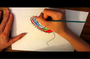
Once the samples have been collected the macroscopic survey will be ready to begin. In order to asses the different samples, petri dishes, vials, pipettes and other lab materials will all be tools of assistance to utilize for better comprehension and overall procedure in the lab. Each one in the group will be responsible to portray to the rest of the team members with their findings in each sample. To do so, we will be drawing whatever is observed through the microscopic view. For the drawings it is key to use a well functioning pencil and a collection of color pencils to better portray shades or variations in coloring, as seen Figure 1.

1

Figure 1. A Drawing of a Bacteria Cell. maxresdefault.jpg

2


Figure 2. Examples of Drawings of Microinvertebrates. Teachengineering.org

The drawings will be done in the lab, and then the group will work together to have a cohesive understanding of the drawings. We envision the most challenging part of the process will be to identify the individual organisms, but an accurate portrayal will be key to surpass this stage in the methods. The drawings will then be compared and analyzed as a group to get an accurate identification of every individual.

Once the individuals have been identified, it will be time to start the written report and the possible conclusions that could be reflected from our findings. Our ultimate goal is, that with the proper identification, we will be able to know which species of micro invertebrates are more commonly seen, and how these species might be affecting the city we live in.  Micro invertebrates are usually forgotten by the inhabitants of every city, even though they undeniably form a part of our every day life, and this is what this study is aiming to stop.

Post by Felipe Gomez


Micro-organisms

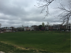
As a team, we decided to individually collect bacteria from meaningful places around campus that are also representative of the types of bacteria that can be found throughout Charlottesville. My everyday walk involves passing through the one and only Madison Bowl, so I decided to make it the source of my bacterial sample. Mad Bowl (as we call it for short) is host to many sports, events, people, pets and, even if we don’t think about it much, it is also hosts to an array of microorganisms, including bacteria. I will collect soil from Mad Bowl, carefully picking soil that is closer to the grass, since bacterial colonies tend to live closer to plant roots. The bacteria in this sample is likely to be representative of the bacteria found specifically on ground soil, given that green spaces around campus get the same maintenance and environmental factors.

We will meet as a team to observe the bacteria of each different sample under a microscope. It will be interesting to see how bacteria differ or are similar to one another between all of our samples.

Finally, since we can’t photograph what we see and using words to describe bacteria can be a dry experience, we decided to record our observations by drawing them. Whether the result is accurate representation or abstract art, I am not sure, but it will certainly be exciting to see what we all come up with! Who thought that documenting bacteria could be art? I certainly didn’t, but stay tuned to see our upcoming “collection”!

5

Even with gloomy weather, we are still mad about Mad Bowl! (Plus, rain means moist soil, and moist soil means optimal environment for bacteria to thrive!)

 Post by Adriana Castillo-Fischer


Microorganisms- Hidden in Plain Sight

Microorganisms are found all over the world. They can reside deep in the ocean, high in the atmosphere, the boiling waters of natural hot springs, and even the frigid grounds of the University of Virginia. For our BioGrounds research project we are focusing on the places where microorganisms are found and what the organisms are doing.

In order to simplify this microscopic survey, we are focusing in on eukaryotic organisms. A eukaryote is a classification of organism that has more than one cell with a nucleus. While humans and other tangible animals, are eukaryotes, many are also microscopic. They usually happen to be the largest type of microscopic organism and will therefore be the focus of our study for a variety of reasons. First, since they are the largest, we will not need the most high tech equipment. This will allow us to spend less time calibrating the equipment and more time searching for the hidden organisms. Second, they will be easier to identify. No one on our team has any extensive biological backgrounds which would make identifying bacteria, archaea, and prokaryotes very difficult. Instead, we will focus on a more well-known group of microorganisms, so that we can quickly identify the species. Third, we are attempting to mimic the works of Agnes Catlow, and her work Drops of Water. In this series, Catlow sketches the microorganisms she sees under the lens of a microscope. Since this book was published in the 19th century, we can assume that she saw mainly eukaryotes and possibly some of the largest protists. For these reasons, we want to focus on cataloging the micro-eukaryotic species around grounds.

Essentially our project is taking her method of capturing the existence of microorganisms and applying it to the grounds of the University of Virginia. By doing so, we hope to catalog a variety of different microorganisms in a variety of places on grounds. In order to begin our project, we need a few research tools. A light microscope, slides, pipettes, petri dishes, and collection vials will all play an important part in collecting and analyzing our data. We are also adding an artistic component to this project and for this we will all use our own preferred method of documentation, but it will most likely included water color, colored pencils, and hand drawing.

We are also trying to get a large sample of what kind of organisms live where on the University’s grounds. We have discussed collecting samples from bicycle tires, bus seats and floors, gutters, sidewalks, dirt, dining halls, and basically any other places that we can. The hope is to find as many microorganisms as possible and document them, to show what a variety we have on grounds. We also want to show that our city is teeming with life, even though we cannot always see it, reinforcing the idea that we live in a biophilic city.

Post by Laura Moran